BME PhD Candidate, Hamed Vavadi, has been awarded a Third Bridge Grant in the amount of $35,000 and first year BME graduate student, Stephanie Knowlton, has been awarded a Third Bridge Grant in the amount of $45,000. This grant aims to identify and provide funding to engineering students and entrepreneurs with promising technologies to advance their technologies towards successful commercialization.
For more on the Third Bridge Grant see the website here
Hamed Vavadi is currently a PhD candidate in the department of Biomedical engineering and he is doing research in Optical and Ultrasound Imaging Laboratories with Professor Quing Zhu to develop imaging systems for detecting cancer and monitoring patients undergoing Neoadjuvant chemotherapy with funding support by National Institute of Health. He is specifically working on combining ultrasound and NIR imaging modalities for clinical diagnosis of breast cancers. His research interest includes, optical imaging, NIR spectroscopy, cancer detection, bio instrumentation and vital signal processing.
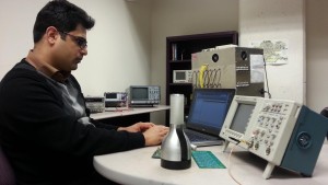
Hamed Vavadi
According to the World Health Organization, breast cancer is the most common cancer among women worldwide, claiming the lives of hundreds of thousands of women each year and affecting countries at all levels of modernization. The National Cancer Institute estimated that each year over 220,000 women in the United States will be diagnosed with breast cancer and more than 40,000 will die. It is well agreed in the medical community that early detection is the most effective way to fight the breast cancer. The current imaging modalities for breast cancer screening and diagnoses such as X-ray mammogram, ultrasound imaging, and magnetic resonance imaging (MRI) are not effective enough to fully address this critical issue and researchers are still looking to find a better solution for early detection of breast cancer. Compared to existing technologies, near infrared (NIR) diffuse optical tomography (DOT) has demonstrated great potential in initial diagnosis of tumor, the assessment of tumor vasculature response to neoadjuvant chemotherapy and distinguishing benign from malignant tumors.
Hamed Vavadi and Prof. Quing Zhu (Optical and ultrasound imaging laboratories) are working on a technology, called ultrasound-guided optical tomography which use near-infrared light to detect complementary functional parameters of breast cancer, such as tumor angiogenesis and hypoxia, noninvasively. This technique has significant potential to assist the characterization of benign and malignant cases and reduce unnecessary normal biopsies. With the support of the Third Bridge Grant they will start exploring the commercialization potential of this technology.
Stephanie Knowlton earned her BS from UConn in 2015 as and is now a graduate student in Biomedical Engineering. Her current research interests include using 3D printing and magnetic levitation to develop medical technologies which can ultimately be applied in clinical settings and enhance healthcare in developing countries. Stephanie is a member of Phi Sigma Rho, a sorority for women in engineering and the Biomedical Engineering Society.
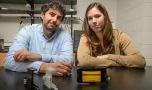
Stephanie Knowlton and her advisor Dr. Savas Tasoglu
Stephanie’s research focuses on the development of a portable diagnostic platform. Currently, many medical diagnostic procedures require advanced, expensive testing equipment, a trained technician, and labor-intensive protocols. This results in delays to patient treatment up to several days, as samples must be sent to remote testing labs where specialized and costly equipment is available. To address this need in the medical field, we have designed a low-cost, automated disease diagnostic platform which will serve as an on-site alternative to sending samples to a remote lab for testing.The compact and portable diagnostic platform functions by levitating a patient’s cells (such as a drop of blood) in a magnetic field, imaging the cells, and performing an automatic analysis on the same device to count cells based on their densities. Based on this analysis, the user interface will display medical diagnostic information for the physician. The technology can be applied to a range of diseases, such as diagnosis of sickle cell anemia. In addition, the device is user-friendly and automated, making it useful for untrained users in clinical settings, in patients’ homes, and in low-resource communities. This technology will allow medical treatment to be administered in a timely manner and enable world-wide access to diagnostic procedures.
Published: Jan 29, 2024